At TheHealthBoard, we're committed to delivering accurate, trustworthy information. Our expert-authored content is rigorously fact-checked and sourced from credible authorities. Discover how we uphold the highest standards in providing you with reliable knowledge.
What are the Different Types of Medical Diagnostic Imaging?
Medical diagnostic imaging refers to technologies that create images of the inside of the body in order to diagnose a medical condition or disease. When doctors assess a patient’s signs and symptoms in order to identify the patient’s condition or disease, the process and conclusion reached is called a medical diagnosis. Because signs and symptoms do not always present themselves on the outside body, doctors use medical diagnostic imaging to check for abnormalities inside the body. Different types of medical diagnostic imaging include X-ray, ultrasound, computed tomography (CT) scans, magnetic resonance imaging (MRI), and nuclear medicine scans. A patient’s experience will depend on what type of technology is used, but most imaging procedures are fast and cause the patient minimal discomfort.
An X-ray, also called Roentgren waves or radiography, is an example of the quick and painless variety of diagnostic imaging. X-rays use electromagnetic radiation to create a picture of the inside of the body in which denser objects, such as bone, are highlighted and less dense objects, such as fat, appear in shades of gray. This technology may be used to determine whether the patient has broken or chipped bones, spinal injuries, bone- or joint-affecting diseases, heart or lung disease, a punctured lung, or scoliosis, the abnormal curving of the spine. X-ray may also be used to locate accidentally swallowed objects, evaluate the cause of chest pain, detect blockages in blood vessels, inspect sinus infections, and evaluate dental problems. X-rays produce a small amount of radiation, produce little discomfort, and are safe for use on children.

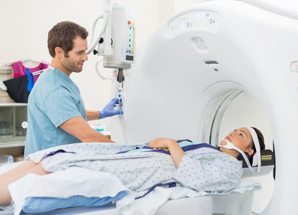
A CT scan, also called a CAT scan, is a high powered type of X-ray technology that looks for internal bleeding, broken bones, blood clots, tumors, musculoskeletal diseases, arterial blockages, and heart disease. During this examination, the patient lies on a table and moved through a donut hole-like opening in the X-ray machine. The patient may also be administered a contrast material to help create a clearer picture. While this test is painless, it may require the patient to remain still inside the machine for several minutes, causing discomfort for those with claustrophobia or chronic pain. Patients should tell their doctor if they may be pregnant.

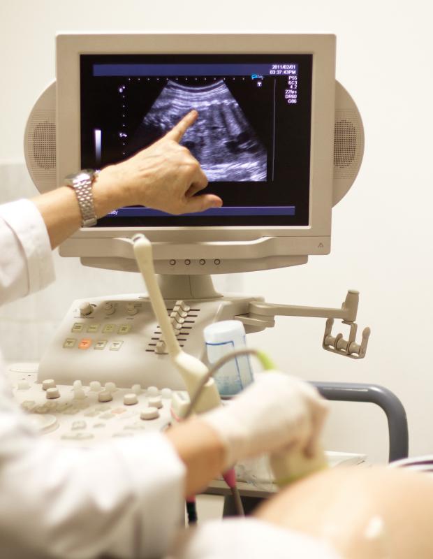
An ultrasound, also called a sonogram, uses sound waves to make a picture of the body’s tissues. They are often used to examine the progress of a fetus in the womb, the heart, blood vessels, kidney, liver, and other internal organs. In this type of medical diagnostic imaging, the doctor applies a warm gel to the outer body overlying the area that will be examined. The physician then moves a device called a transducer, which produces high frequency sound waves, over the body. The sound waves bounce off tissues and the transducer collects the returning waves and creates a picture of the organs. Ultrasound is safe, painless, and produces no radiation, but it is not effective at imaging areas of the body that contain gas or are located beneath bone.

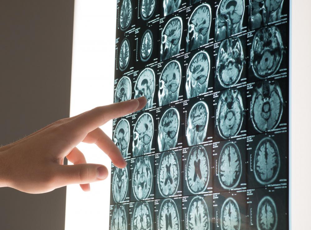
MRIs are a type of medical diagnostic imaging used to look at the spinal cord, brain, heart, blood vessels, and other internal organs. The patient lies as still as possible within the MRI machine while the machine uses magnet and radio waves to make a three-dimensional picture of the inside of the body. Most patients will not experience any discomfort, unless they have trouble being in an enclosed space or staying still. The test usually has no harmful effects, but patients should let their doctor know if they may be pregnant, have any electronic devices, such as a pacemaker, inside them, or any pieces of metal in their body.

Nuclear scans, also called radioisotope scans or radionuclide scans, use a radioactive substance and a gamma ray camera to make a picture of the inside of the body. The radioactive substance is administered in a very small, harmless dosage either orally or intravenously, through injection, and allowed to travel through the body. When the substance reaches the part of the body needing to be examined, the patient lies in a machine for twenty to thirty minutes. The two dimensional pictures taken by the machine will show whether the tissue is functioning properly or not.
AS FEATURED ON:
AS FEATURED ON:

-
![A magnetic resonance imaging scanner.]() By: Andrey NavrotskiyA magnetic resonance imaging scanner.
By: Andrey NavrotskiyA magnetic resonance imaging scanner. -
![Ultrasound is a type of medical diagnostic imaging system that is used to monitor pregnancies.]() By: Konstantin LiUltrasound is a type of medical diagnostic imaging system that is used to monitor pregnancies.
By: Konstantin LiUltrasound is a type of medical diagnostic imaging system that is used to monitor pregnancies. -
![Medical diagnostic imaging often allows doctors to make a diagnosis without invasive testing.]() By: michaeljungMedical diagnostic imaging often allows doctors to make a diagnosis without invasive testing.
By: michaeljungMedical diagnostic imaging often allows doctors to make a diagnosis without invasive testing. -
![X-rays, ultrasonds and computed tomography scans are among commonly used diagnostic imaging technologies.]() By: adam121X-rays, ultrasonds and computed tomography scans are among commonly used diagnostic imaging technologies.
By: adam121X-rays, ultrasonds and computed tomography scans are among commonly used diagnostic imaging technologies. -
![Dentists may use several different types of X-rays to get a full picture of the situation below the gum line.]() By: Robert KneschkeDentists may use several different types of X-rays to get a full picture of the situation below the gum line.
By: Robert KneschkeDentists may use several different types of X-rays to get a full picture of the situation below the gum line. -
![A CT scan combines a rotating X-ray beam with computer technology to create cross-sectional images of the body.]() By: Tyler OlsonA CT scan combines a rotating X-ray beam with computer technology to create cross-sectional images of the body.
By: Tyler OlsonA CT scan combines a rotating X-ray beam with computer technology to create cross-sectional images of the body.


Discuss this Article
Post your comments