At TheHealthBoard, we're committed to delivering accurate, trustworthy information. Our expert-authored content is rigorously fact-checked and sourced from credible authorities. Discover how we uphold the highest standards in providing you with reliable knowledge.
What is Cardiac Ultrasound?
 Mary McMahon
Mary McMahon
Mary McMahon
Cardiac ultrasound or echocardiography is a medical imaging procedure in which the goal is to generate a picture of the heart for the purpose of evaluating a heart condition or suspected heart problem. Like other types of ultrasound imaging, cardiac ultrasound is non-invasive and painless, and it can be performed as an outpatient procedure in a clinic or hospital. There are a variety of reasons for a doctor to request a cardiac ultrasound, and he or she will usually discuss the reason for the procedure with the patient at the time that the procedure is recommended.
Ultrasound imaging relies on the use of high frequency sound waves to create a picture of the inside of the body. The waves are emitted by a transducer which also picks up the waves when they bounce back, generating a picture on the basis of the way in which the sound waves change as they bounce off objects in the chest cavity. In a basic ultrasound, a flat picture of the heart is produced, and it is also possible to generate a three dimensional ultrasound image with the use of an array of transducers. Both still images and video can be captured with echocardiography.

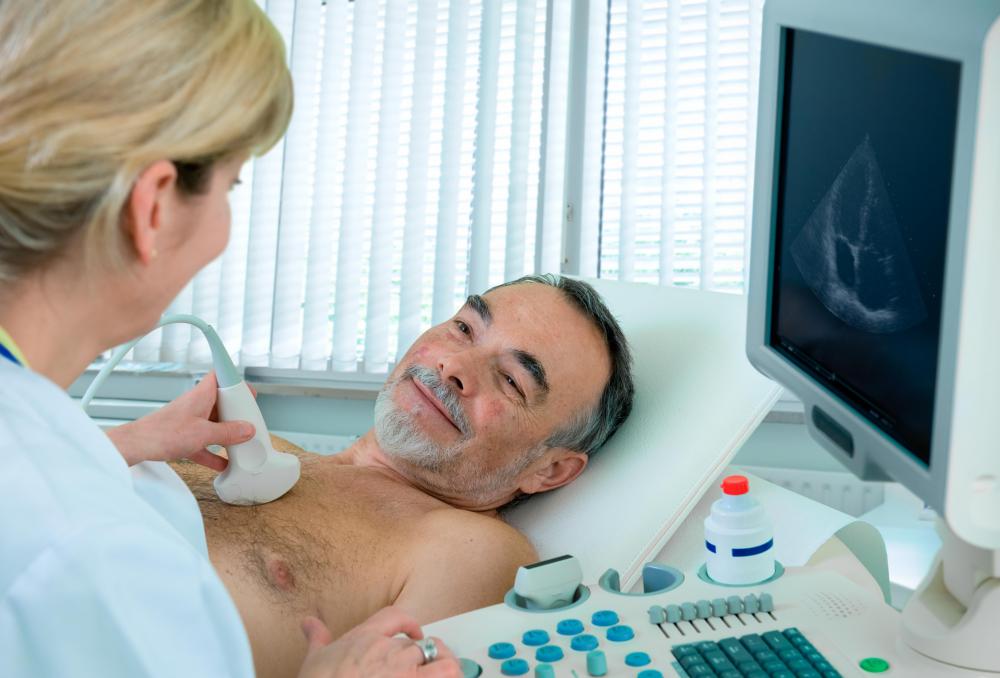
In a cardiac ultrasound, the patient's chest will be exposed and covered in a conductive gel. The ultrasound technician or doctor will move the transducer across the chest, occasionally changing angles to get a better picture. The patient must hold still for a clear picture, and the patient may need to hold his or her breath at certain points to avoid movements in the chest which could obscure the ultrasound image.

Patients usually lie down or are propped in a partially seated position for this ultrasound. In some cases, the doctor may ask the patient to exercise before a cardiac ultrasound so an image of the heart under stress can be produced. In this case, an image of the resting heart may be acquired before the heart is stressed.
If a doctor performs the ultrasound, he or she may discuss the ramifications of the results immediately with the patient. In other instances, the doctor may want to review the ultrasound images along with the results of bloodwork and other tests before discussing the patient's situation. Ultrasound technicians are usually not allowed to discuss medical information with patients, instead turning the images over to a radiologist or doctor for interpretation.
Mary McMahon
Ever since she began contributing to the site several years ago, Mary has embraced the exciting challenge of being a TheHealthBoard researcher and writer. Mary has a liberal arts degree from Goddard College and spends her free time reading, cooking, and exploring the great outdoors.
Learn more...
Mary McMahon
Ever since she began contributing to the site several years ago, Mary has embraced the exciting challenge of being a TheHealthBoard researcher and writer. Mary has a liberal arts degree from Goddard College and spends her free time reading, cooking, and exploring the great outdoors.
Learn more...AS FEATURED ON:
AS FEATURED ON:

-
![A doctor may ask a patient to exercise prior to a cardiac ultrasound.]() By: Alexander RathsA doctor may ask a patient to exercise prior to a cardiac ultrasound.
By: Alexander RathsA doctor may ask a patient to exercise prior to a cardiac ultrasound. -
![Cardiac ultrasounds use sound waves to monitor the condition of a patient's organs.]() By: Alexander RathsCardiac ultrasounds use sound waves to monitor the condition of a patient's organs.
By: Alexander RathsCardiac ultrasounds use sound waves to monitor the condition of a patient's organs. -
![Before an ultrasound, a small layer of conductive gel is applied to make the images clearer.]() By: Klaus EppeleBefore an ultrasound, a small layer of conductive gel is applied to make the images clearer.
By: Klaus EppeleBefore an ultrasound, a small layer of conductive gel is applied to make the images clearer. -
![It's possible to generate a three-dimensional ultrasound image of the heart with an ultrasound.]() By: Vladislav GajicIt's possible to generate a three-dimensional ultrasound image of the heart with an ultrasound.
By: Vladislav GajicIt's possible to generate a three-dimensional ultrasound image of the heart with an ultrasound.


Discussion Comments
I recently had an echo exam and since then I've had symptoms of an allergic reaction. Has anyone experienced this? My skin itches, but it's not severe. Also I have a slight headache, and a raspy feeling in my throat. I have not eaten or used anything different recently.
I think it's amazing they can use ultrasounds to make a 3-D picture of your heart! Instead of having to do an invasive exploratory surgery to see what it looks like, the doctor can just do a cardiovascular ultrasound to see what is going on.
I also think it's cool that this kind of medical imaging doesn't require the use of radiation. I know that being exposed to a little bit of radiation for an x-ray won't really hurt you, but it's not very good for the technicians. It seems like ultrasound technicians don't have to worry about this!
@JaneAir - I actually have a friend from college who works as a medical sonographer. She told me that when she went to school, she was able to choose a specialty, and there was a track specific for people who wanted to specialize in cardiac ultrasounds. My friend decided just to do the general track, but a good number of her classmates chose the cardiac track.
I think it's good that ultrasounds are a popular kind of medical imaging, because they are totally painless. I had an abdominal ultrasound a few years ago, and i didn't feel a thing. The only slight annoyance was that the gel they had to put on my stomach was kind of cold, but that was easy to deal with.
I was actually reading another article about cardiac ultrasounds the other day! Apparently ultrasounds for stuff besides pregnancy are growing more and more popular in the medical community. They're especially good for cardiac stuff because they are non-invasive, but allow the doctor to see what's going on.
Anyway, the article said that there is a growing need for ultrasound technicians because of this upswing in ultrasound use. So if you're looking to change careers and get into the medical field, you might consider looking into that.
what is the diagnostic technique to detect the calcium score in coronary arteries?
Would this be the same diagnostic technique to detect hypertrophic cardiomyopathy (HCM)?
Post your comments